Unlock up to 10,000 USDT with the best MEXC referral code, mexc-NFTP. Learn how to claim bonuses, reduce trading fees, and earn rewards for deposits and trades.
Scientists use Lumia 1020 to sequence DNA on the cheap

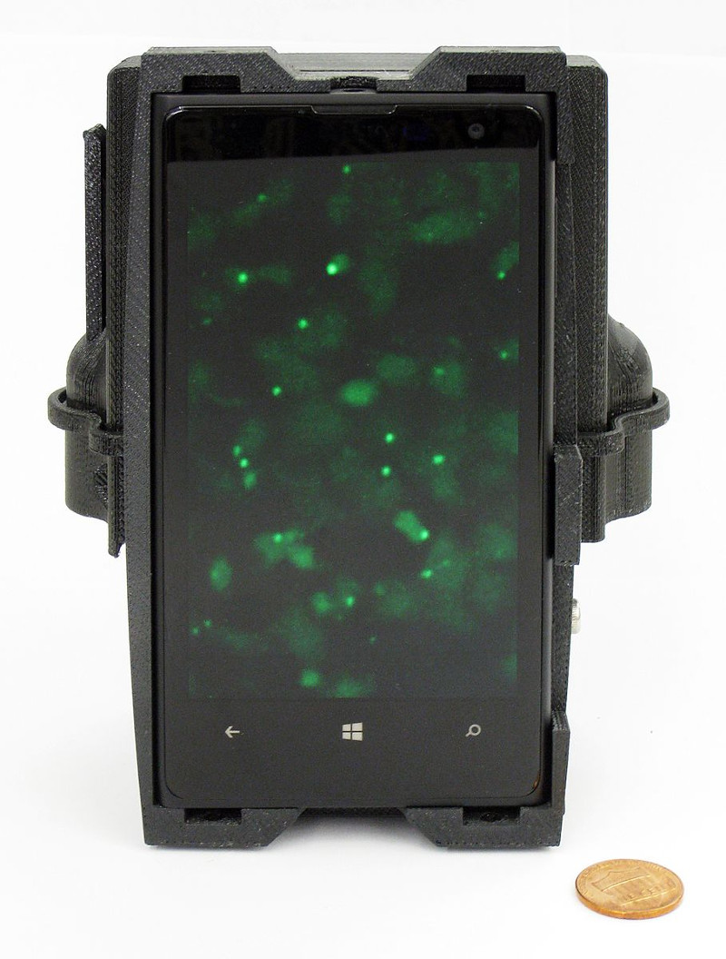
Scientists from UCLA and Sweden have successfully sequenced DNA using a modified Lumia 1020 smartphone.
In a press statement, the institution said that DNA sequencing typically sees samples being sent to well-equipped facilities far away, representing a challenge for developing countries and underserved communities.
But the smartphone-based microscope, developed by researchers at UCLA’s California NanoSystems Institute and scientists from Stockholm University and Uppsala University, could reduce the need for expensive equipment.
The device takes the form of a lightweight, 3D-printed optical attachment, fitted to the back of a Lumia 1020 and used in conjunction with the 41-megapixel camera.
“The device can image and analyse specific DNA sequences and genetic mutations in tumor cells and tissue samples without having to first extract DNA from them,” UCLA explained.
Microsoft’s Lumia 1020 still stands out thanks to its gargantuan 41MP main camera – and scientists are putting it to good use
Aydogan Ozcan, one of the lead researchers, says that the attachment could be manufactured for less than US$500.
“A typical microscope with multiple imaging modes would cost around $10 000, whereas higher-end versions, such as the one we used to validate our mobile-phone microscope, would go for $50 000 or more,” he adds.
Using the attachment is a pretty seamless task too, it seems.
“To use the device, a technician places a tissue sample in a small container. The mobile phone microscope records multi-mode images of the processed sample and feeds data to an algorithm, which automatically analyses the images to read the sequenced DNA bases of the extracted tumor DNA, or to find genetic mutations directly inside the tumor tissue. This mobile microscope can detect even small amounts of cancer cells among a large group of normal cells.”
Featured image: Karlis Dambrans via Flickr


